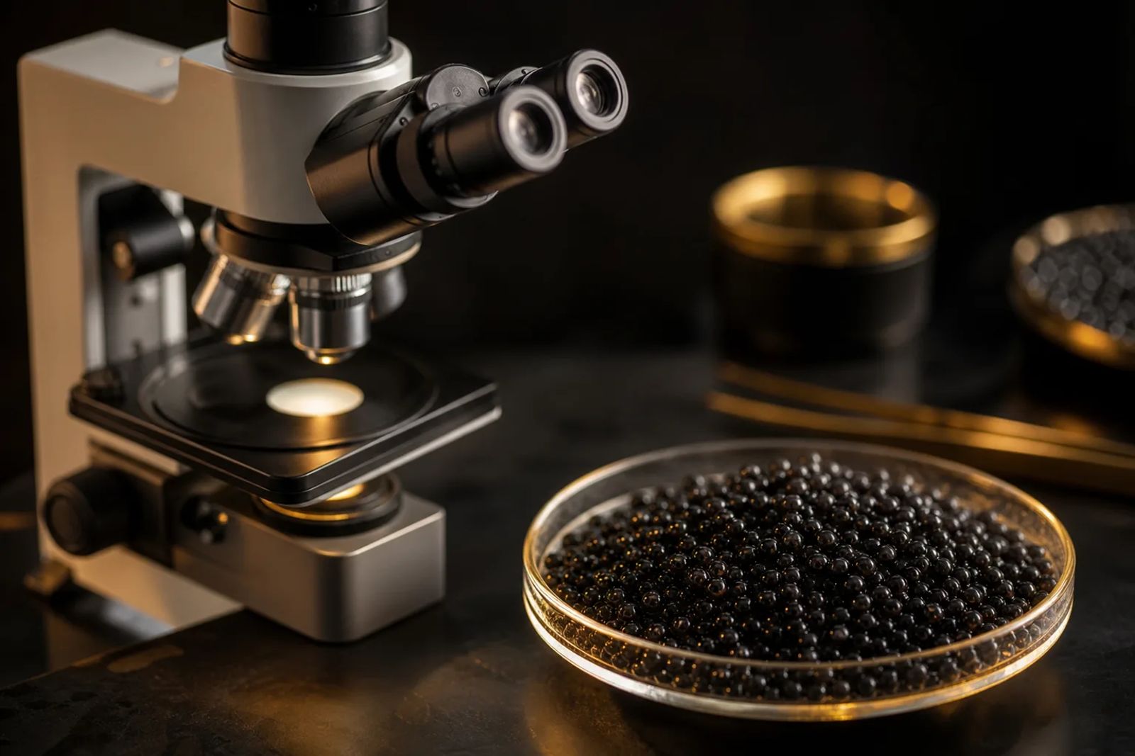
Inside a sturgeon ovary, a follicle can break down and its contents be resorbed instead of producing an ovulated egg. Linares-Casenave and colleagues documented this process in a 2002 white sturgeon experiment. This is atresia: the degeneration and resorption of follicles, not another step towards ripeness.
Healthy development follows a different course: oocyte growth, yolk deposition, final maturation and ovulation. Traditional caviar is made from ovarian oocytes collected before ovulation. Understanding that sequence reveals how the structures needed for intact caviar beads are built, and how they can be lost.
Which oocyte staging scale is being used?
A stage number needs a named scale. Here, the reference is Serge Doroshov’s five-stage white sturgeon scheme, reported in California Sea Grant’s 1994 research report on ovarian development. It separates resting follicles, endogenous growth, previtellogenesis, exogenous vitellogenesis and postvitellogenic polarisation. Another paper’s ovarian maturity scale may assign its numbers differently.
The terms matter too. An oocyte is the developing egg cell; a follicle includes that cell and its surrounding support tissues. An oocyte stage therefore describes something more specific than the condition of the whole ovary. For the wider anatomy, see sturgeon biology.
| Stage | Defining features | Detection | Relevance to caviar | Source |
|---|---|---|---|---|
| I, resting | Quiescent follicle | Histology | Not a harvest stage | Doroshov, 1994 |
| II, endogenous growth | Growth before external yolk deposition | Histology | Not a harvest stage | Doroshov, 1994 |
| III, previtellogenic | Chorion synthesis; granulosa-cell proliferation | Histology | Envelope formation, not ripeness | Doroshov, 1994 |
| IV, vitellogenic | Exogenous yolk accumulation | Histology; follicle measurements | Growth towards harvestable roe | Doroshov, 1994 |
| V, postvitellogenic | Yolk growth complete; nucleus polarises | Sectioned oocytes; polarisation index | Tracks development within full-sized oocytes | Doroshov, 1994 |
| Final maturation and ovulation, outside this scale | Meiosis resumes; follicle releases egg | Maturation assays; observation | Ovulated eggs differ from traditional roe | Chapman and Van Eenennaam, 2016 |
| Atresia, outside this scale | Follicle degeneration and resorption | Histology; supporting blood measurements | Roe structures damaged | Talbott et al., 2011 |
Vitellogenesis: building the yolk reserve
A growing oocyte is stocking a reserve for the embryo. Vitellogenesis fills it with yolk, which supports development before external feeding begins. Vitellogenin, a yolk precursor produced principally in the liver, travels through the blood to growing oocytes. This is active nutrient provisioning, rather than simply an egg taking up water.
In captive sterlet, a study of gene expression and biochemical indicators found that vitellogenin expression increased towards late vitellogenesis in both the liver and ovary.
Building that reserve takes time. Linares-Casenave and colleagues’ 1997 conference abstract reported vitellogenesis lasting 16 to 18 months in domestic white sturgeon. That timescale belongs to those fish and rearing conditions; it neither applies to every sturgeon nor covers the whole interval from hatching to a female’s first caviar harvest.
Diameter tells how much an oocyte has grown. Tissue examination reveals what it contains. Together, these observations help distinguish a growing vitellogenic follicle from a full-grown follicle whose internal maturation is advancing. Record species, individual identity and thermal history alongside the measurement. A calendar date alone cannot identify a developmental stage.
Germinal vesicle migration and the polarisation index
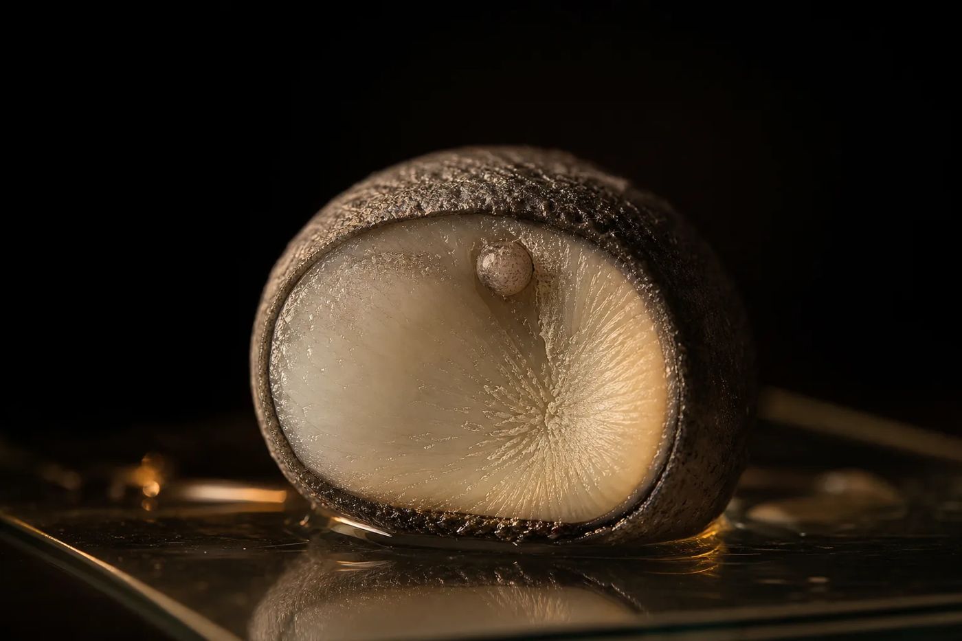
As late development advances, the oocyte’s nucleus moves towards its edge. This nucleus is the germinal vesicle, or GV, and its destination is the animal pole, the end associated with subsequent nuclear maturation.
The polarisation index, or PI, expresses the GV’s remaining distance from the cell surface relative to oocyte diameter. A smaller value places the GV closer to the periphery.
PI = B/A, where B is the distance from the GV’s outer edge nearest the animal pole to the oocyte plasma membrane, and A is the oocyte diameter along the animal-vegetal axis, excluding the chorion. Chapman and Van Eenennaam illustrate this definition in UF/IFAS publication FA153.
Measuring it demands a precise cut. FAO’s Sturgeon Hatchery Manual describes fixing sampled oocytes, bisecting them along that axis, then measuring the section with a binocular microscope and eyepiece micrometer. Cut obliquely, miss the GV or include the envelope, and the measurement changes even though the biology has not. Report the preparation method and the number of oocytes examined alongside the PI.
PI measures nuclear position. It does not grade flavour or texture, or diagnose atresia on its own. Numerical criteria for spawning induction apply to the protocols and populations in which they were evaluated, rather than serving as established caviar harvest limits. For their practical use in farm decisions, see checking egg ripeness.
Final maturation, ovulation and the chorion
A full-grown egg has not necessarily completed maturation. During final oocyte maturation, meiosis resumes and the GV envelope breaks down. Meiosis is the specialised cell division that prepares the maternal chromosomes. Germinal vesicle breakdown, or GVBD, is a nuclear event; ovulation is the subsequent release of the egg from its follicle.
Traditional caviar uses preovulatory ovarian oocytes, not eggs already released for spawning. The distinction includes the surrounding follicular tissues, rather than simply the harvesting method used to remove the eggs. Ovulated eggs need separate handling considerations, explored in no-kill caviar and ovulated eggs.
The chorion is the extracellular egg envelope. It is distinct from the living plasma membrane and from the outer follicular cells. Doroshov’s histological scheme records its synthesis before exogenous vitellogenesis begins. Its mechanical role is more complex than a single explanation based on membrane thickness; see the egg membrane and caviar texture.
Atresia: degeneration rather than ripening
Atresia dismantles what development has built. Instead of producing an ovulated egg, the follicle breaks down and its contents are resorbed. Alongside digestion of the egg envelope and disruption of the oocyte’s outer cytoplasm, Linares-Casenave and colleagues’ 2002 white sturgeon experiment documented enlarged granulosa cells. Such damage may compromise bead integrity, although the study did not measure processed caviar texture.
Temperature has direct experimental support as a trigger. The researchers exposed females late in oogenesis to warm water at 18 to 20 °C (64 to 68 °F), or to seasonal control temperatures of 12 to 16 °C (54 to 61 °F). Among warm-exposed females, those that had not acquired maturational competence showed arrested development followed by atresia. These findings concern white sturgeon at that developmental transition, rather than establishing a damaging temperature for all sturgeons.
Handling and transport stress can affect reproductive success. The cited studies do not establish whether they directly cause atresia on farms, so stress and atresia remain distinct concerns.
Cut obliquely, miss the GV or include the envelope, and the measurement changes even though the biology has not.
Timing also matters when advanced females are held beyond their suitable physiological window. FAO’s manual warns of resorption after prolonged holding of ripe females at spawning temperature. This broodstock-management observation does not set a maximum waiting time for every caviar farm. The role of environmental management is explored in water temperature, light and maturation.
Detecting early atresia
Early atresia can escape gross inspection. Histology reveals tissue changes directly; blood markers provide supporting indicators.
In Talbott and colleagues’ 2011 study, ten farmed white sturgeon underwent repeated blood sampling and ovarian biopsies while transfer from cold to warm water induced atresia. Testosterone concentrations were lower when early histological signs appeared. A testosterone-based discriminant analysis classified normal and atretic fish with high accuracy within that dataset.
The finding comes from a small, induced-atresia experiment, rather than validation across species or farms. For instrument choice and sampling approaches, see sexing and examining sturgeon.
What stage can tell us about caviar quality
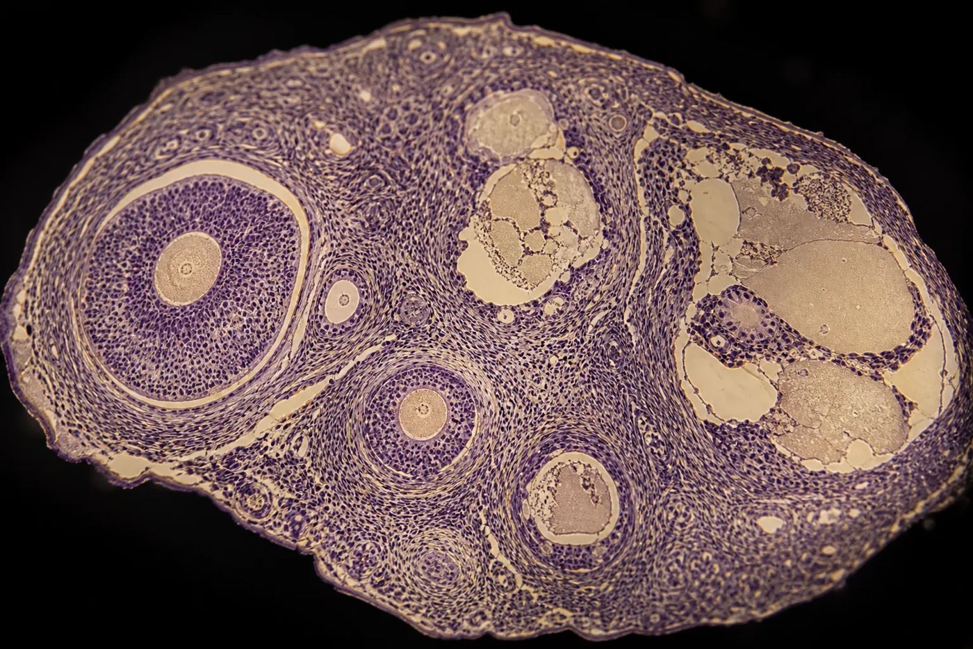
The clearest connection is structural. Developing follicles build the egg’s reserves and coverings; atresia destroys them. Chapman and Van Eenennaam describe oocytes softening and changing shape as ovulation approaches, and losing firmness with resorption. Their extension guidance explains the practical concern, without supplying a controlled sensory trial that establishes an optimum PI.
A useful counterweight to that structural explanation comes from Yue Zhang’s 2011 University of California, Davis, thesis on husbandry and diet. It followed 92 individually marked white sturgeon females across two farms, with dietary treatments followed by cold-water holding. At harvest, measurements included egg diameter, PI and egg texture. Farm, diet and their interaction did not significantly affect those endpoints in the reported analysis.
That result describes the conditions studied, rather than ruling out husbandry effects on quality. The essential distinction is between a plausible biological explanation and an effect demonstrated under particular conditions. Evidence about fertilisation or embryo survival likewise answers a different question from evidence about the texture of salted caviar.
Sampling adds another consideration: how well does one sample represent an ovary? Talbott and colleagues’ 2014 study compared PI and follicle diameter at three positions per ovary in five postvitellogenic white sturgeon. Some differences were statistically detectable but judged biologically unimportant. That supports local sampling for that population and stage, without establishing uniformity in every developing or regressing ovary.
For farm records, keep four observations separate: growth stage, nuclear position, follicle integrity and the measured properties of processed roe. Read together, they distinguish development, impending ovulation and degeneration. Each contributes something useful; no single measurement can promise caviar quality.
Sources
-
California Sea Grant, University of California. Doroshov, S., Environmental Effects on Ovarian Development in White Sturgeon Broodstock, in Biennial Report of Completed Projects 1990 to 1992, publication R-CSGCP-038, 1994. Report PDF.
-
University of Florida, IFAS Extension. Chapman, F. A., and Van Eenennaam, J. P., Sturgeon Aquaculture: Specialized Techniques: Determining the Stage of Sexual Maturity in Female Sturgeon for Artificial Spawning: The Egg Polarization Index or PI, FA153, first published 2007, reviewed August 2025. Publication.
-
FAO. Chebanov, M. S., and Galich, E. V., Sturgeon Hatchery Manual, FAO Fisheries and Aquaculture Technical Paper 558, 2013. Manual PDF.
-
Fish Physiology and Biochemistry. Changes of vitellogenin and Lipase in captive Sterlet sturgeon Acipenser ruthenus females during previtellogenesis to early atresia, 2016. Retrieved abstract and bibliographic record. PubMed record.
-
Linares-Casenave, J., Van Eenennaam, J. P., and Doroshov, S. I. Temperature-Induced Ovarian Atresia in the White Sturgeon, Acipenser transmontanus, conference abstract, Third International Symposium on Sturgeon, Piacenza, 1997, reproduced in the Québec Ministry of Environment and Wildlife mission report. Archived report PDF.
-
Wiley, Journal of Applied Ichthyology. Linares-Casenave, J., Van Eenennaam, J. P., and Doroshov, S. I., Ultrastructural and histological observations on temperature-induced follicular ovarian atresia in the white sturgeon, volume 18, pages 382 to 390, 2002. Publisher record and author-uploaded abstract retrieved. Publisher record.
-
Elsevier, Aquaculture. Talbott, M. J., and colleagues, Investigating the use of plasma testosterone and estradiol-17β to detect ovarian follicular atresia in farmed white sturgeon, Acipenser transmontanus, volume 315, pages 283 to 289, 2011. Author-copy PDF.
-
University of Florida, IFAS Extension. Chapman, F. A., and Van Eenennaam, J. P., Technically Speaking, What Is Sturgeon Caviar?, FA194, first published 2016, reviewed February 2025. Publication PDF.
-
University of California, Davis. Zhang, Y., Husbandry and Dietary Effects on Sturgeon (Acipenser transmontanus) Farmed for Caviar, thesis, 2011. Retrieved repository abstract. Repository record.
-
Fish Physiology and Biochemistry. Talbott, M. J., and colleagues, Confirmation of ovarian homogeneity in post-vitellogenic cultured white sturgeon, Acipenser transmontanus, 2014, published online in 2013. Retrieved abstract and bibliographic record. PubMed record.
The piece was researched from primary research reports, retrieved study abstracts, an author-copy paper, FAO technical guidance and university extension publications, keeping experimental findings separate from practical interpretation.
Last checked: November 2026